Small Intestine Function, Anatomy, Location, Length and Diagram
The small intestine is part of your digestive system. It makes up part of the long pathway that food takes through your body, called the gastrointestinal (GI) tract. When food leaves your stomach, it enters the small intestine, also called the small bowel. The small bowel connects to the large bowel, also called the large intestine or colon.
Small Intestine Photograph by Asklepios Medical Atlas Pixels
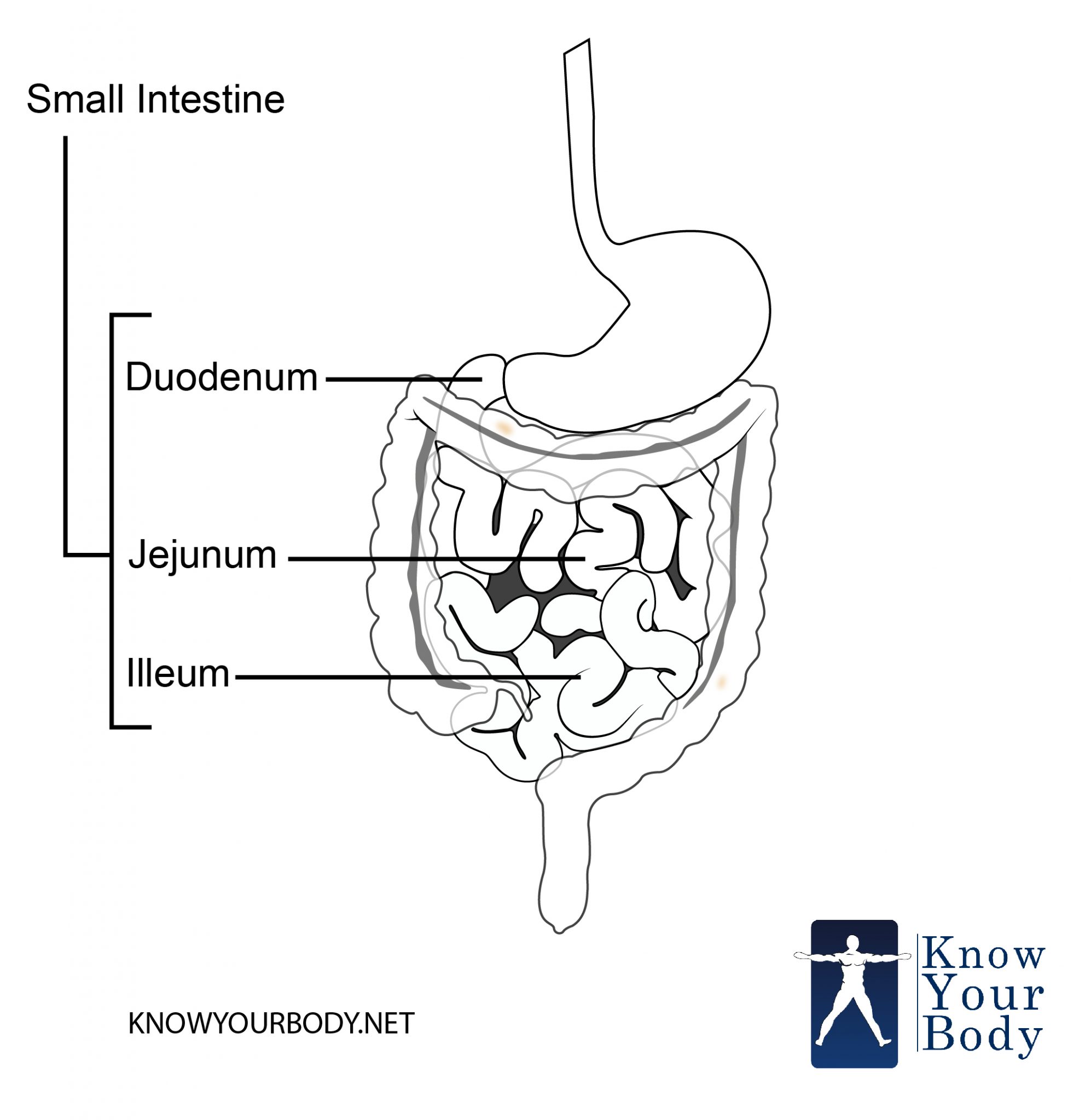
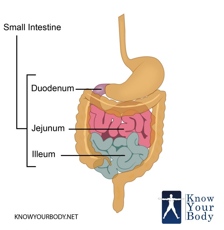
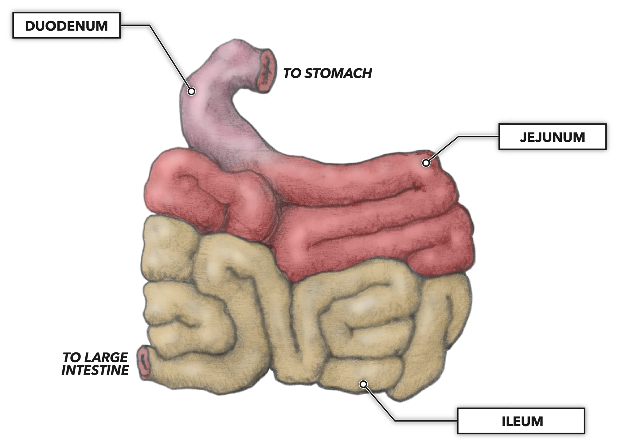
1/4 Synonyms: none The small intestine is the longest part of the digestive system. It extends from the stomach ( pylorus) to the large intestine ( cecum) and consists of three parts: duodenum, jejunum and ileum. The main functions of the small intestine are to complete digestion of food and to absorb nutrients.

Small and large intestine labeled Large intestine, Anatomy, Colon
small intestine, a long, narrow, folded or coiled tube extending from the stomach to the large intestine; it is the region where most digestion and absorption of food takes place. It is about 6.7 to 7.6 metres (22 to 25 feet) long, highly convoluted, and contained in the central and lower abdominal cavity.

small intestine histology labeled Google Search Human anatomy and
The small intestine is made up of the duodenum, jejunum, and ileum. Together with the esophagus, large intestine, and the stomach, it forms the gastrointestinal tract. In living humans, the.
Small Intestine Function, Anatomy, Location, Length and Diagram
marinamedicine. Accounting Final (Elliot) CHAPTER 1 - 8. 28 terms. ethanwillis24. 1 / 4. Study with Quizlet and memorize flashcards containing terms like Duodenum, Jejunum, Ileum and more.

small intestine villi model Google Search
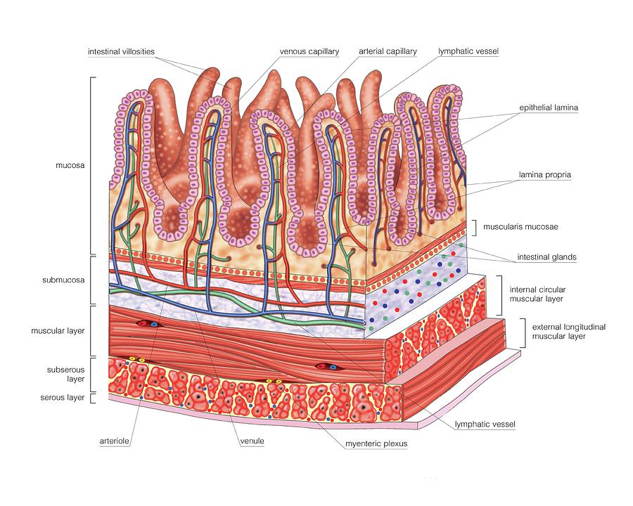
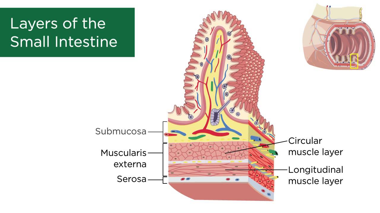
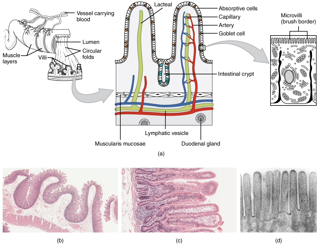
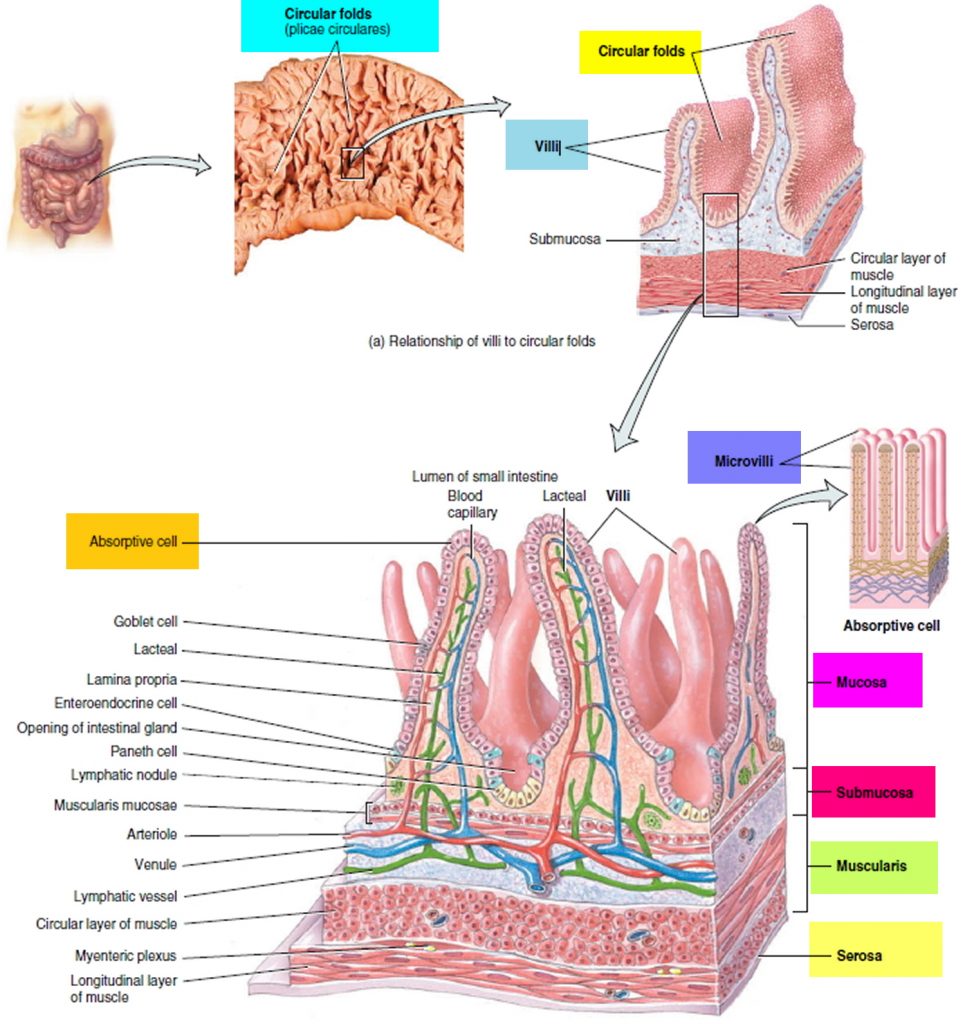
1 Click the card to flip 👆 1 / 11 Flashcards Learn Test Match Created by Terms in this set (11) villus columnar epithelium intestinal crypt 3 muscularis mucosae 4 circular muscle layer 5 longitudinal muscle layer 6 lacteal 7 mucosa 8 submucosa 9 muscularis externa 10 Students also viewed model of villi in duodenum of small intestine 17 terms Images
Layers of the Small Intestine YouTube
The small intestine is a crucial component of the digestive system that allows for the breakdown and absorption of important nutrients that permits the body to function at its peak performance. The small intestine accomplishes this via a complex network of blood vessels, nerves, and muscles that work together to achieve this task. It is a massive organ that has an average length of 3 to 5.

Abdominal Cavity (no liver, stomach, small intestine) model STEM
The Small Intestine's Layers. Section of duodenum: This image shows the layers of the duodenum: the serosa, muscularis, submucosa, and mucosa. The small intestine has four tissue layers: The serosa is the outermost layer of the intestine. The serosa is a smooth membrane consisting of a thin layer of cells that secrete serous fluid, and a thin.

The Small Intestine Complete Anatomy
Content:Introduction 0:00Small Intestine Overview: 00:40Duodenum: 01:14Jejunum and Ileum: 03:39Anatomical Structures of the Duodenum: 6:48Anatomical Structur.

small intestine villi model Google Search Label image, Anatomy
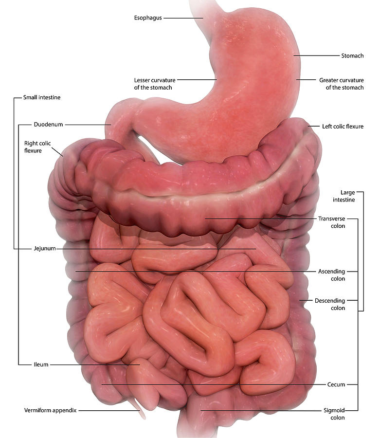
The digestive system. Diseases or Conditions Digestive Diseases File Size 361 KB | 1380 x 1716 File Type JPG The digestive system with sections labeled: mouth, esophagus, liver, stomach, gallbladder, pancreas, small intestine, large intestine, rectum, and anus.
The Small and Large Intestines · Anatomy and Physiology
The small intestine has three distinct regions - the duodenum, jejunum, and ileum.. The primary function of the small intestine is the absorption of nutri.
Small Intestine Location, Function, Length and Parts of the Small Intestine
The mesentery of the small intestine is a large and broad fan-shaped mesentery that is attached to the jejunum and ileum of the small intestine, connecting them to the posterior abdominal wall. Superiorly, the mesentery of the small intestine is attached to the end of the duodenum/beginning of the jejunum (duodenojejunal junction) just to the left of the 2nd lumbar vertebra.

Image result for digestive system models labeled Digestive system
CCC Online Biology Lab - small intestine histology model labeled and unlabeled. Dr. E 421 subscribers Subscribe 132 views 2 years ago Digestive System Silent photo tour of the small.
CrossFit The Gastrointestinal System Small Intestine
The small intestine is an organ located within the gastrointestinal tract.It is approximately 6.5m in the average person and assists in the digestion and absorption of ingested food. It extends from the pylorus of the stomach to the ileocaecal junction, where it meets the large intestine at the ileocaecal valve.Anatomically, the small bowel can be divided into three parts: the duodenum.

CCC Online Biology Lab small intestine histology model labeled and
The small intestine is an organ located in the gastrointestinal tract, between the stomach and the large intestine. It is on average 23ft long and is comprised of three structural parts; the duodenum, jejenum and ileum. Functionally, the small intestine is chiefly involved in the digestion and absorption of nutrients. It receives pancreatic secretions and bile through the hepatopancreatic duct.
Small Intestine Anatomy Labeled Diagramaica
Anatomy (Macroscopy) The small intestine is a specialized tubular structure within the abdominal cavity in continuity with the stomach proximally and the colon distally. The small bowel is about 6 m in the adult. Three subdivisions; "the duodenum, jejunum, and ileum"are defined and characterized by various anatomic relationships.
